Information:
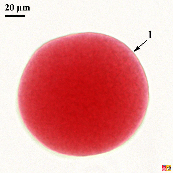
The unfertilized egg is surrounded by a vitelline membrane (= yolk membrane). The yolk material is distributed evenly over the cytoplasm. The sea urchin egg is used as a model to study the cleavage in an isolecithal type of egg (iso = even; lecithal = yolk).
Stained microscopical slide of the unfertilized egg 1 Vitelline membrane
Fertilized egg
Information:
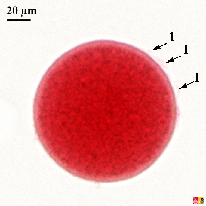
The fertilized egg can be recognized by the presence of a fertilization membrane. This membrane arises immediately after the penetration of a sperm cell and prevents further attempts of fertilization by other sperm cells. The space between the membrane and the yolk is called the perivitelline area. (Both structures are not clearly visible here, but remnants of the fertilization membrane are indicated with 1).
The wonderful video hereabove from YouTube originates from: Essential Cell Biology, 3rd Edition, Alberts, Bray, Hopkin, Johnson, Lewis, Raff, Roberts, & Walter, ISBN: 978-0-8153-4129-1
Stained microscopical slide of the fertilized egg
1 Remnants of the fertilization membrane
Two-cell stage
Information:
The fertilized egg undergoes a complete cleavage, known as holoblastic cleavage (holo = entire; blasto = yolk), by division of the yolk into two equal blastomeres (embryonal cells).
First cell division and formation opf two daughter cells by constriction. Movie made by journalists at the 2009 MBL Logan Science Journalism Program in Woods Hole.
Stained microscopical preparation of the two-cells stage 1 Blastomere (one of the two daughter cells), 2 Blastomere (the other of the two daughter cells), 3 Fertilization membrane, 4 First cleavage plane
Four-cell stage
Information:
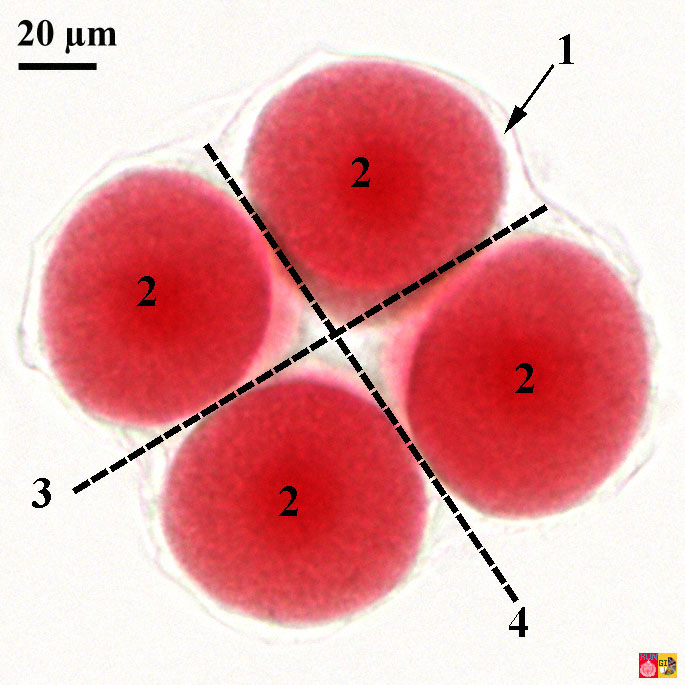
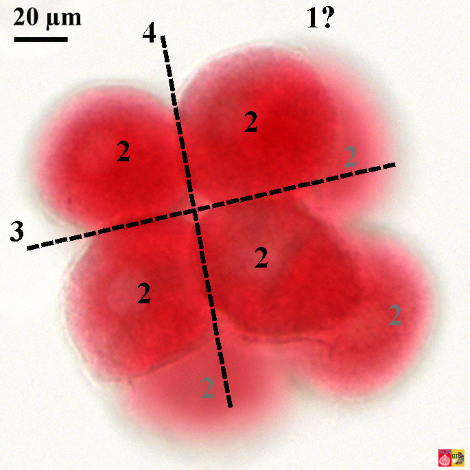
The first and second cleavages are both meridional and are perpendicular (orthogonal) to each other in this isolecithal type of egg.
Diagram of the three-dimensional orientation of the first (1), second (2) and third (3) division plane"
Stained microscopical preparation of the four cells stage 1 Fertilization membrane, 2 Blastomeres (4x), 3 First cleavage plane, 4 Second cleavage plane
Eight-cell stage
Information:
The third cleavage occurs in an equatorial plane, perpendicular to the first two cleavages planes (orthogonal). Here this third division plane in in the same field as the screen. The yolk is more or less equally divided over the blastomeres.
Stained microscopical preparation of the eight cells stage 1 ? Fertilization membrane (not visible here, see four cells stage), 2 Blastomeres (8 x in total), 3 First cleavage plane, 4 Second cleavage plane
Sixteen-cell stage
Information:
The fourth cleavage is very different from the first three. From this point on the orientation of the cleavage planes becomes more and more asymmetrical, leading to the formation of small and larger blastomeres. On account of the cell size, one can distinguish micro-, macro- and mesomeres. The 4 micromeres (mi; small) are located at the vegetal pole (v), the 8 mesomeres (me; medium) at the animal pole (a) and the 4 macromeren (ma; large) in between.
Stained microscopical preparation of the sixteen cells stage 1 ? Fertilization membrane (not visible here, see four cells stage), 2 Blastomeres (16x in total), 3 First cleavage plane, 4 Second cleavage plane
Thirty two-cell stage
Information:
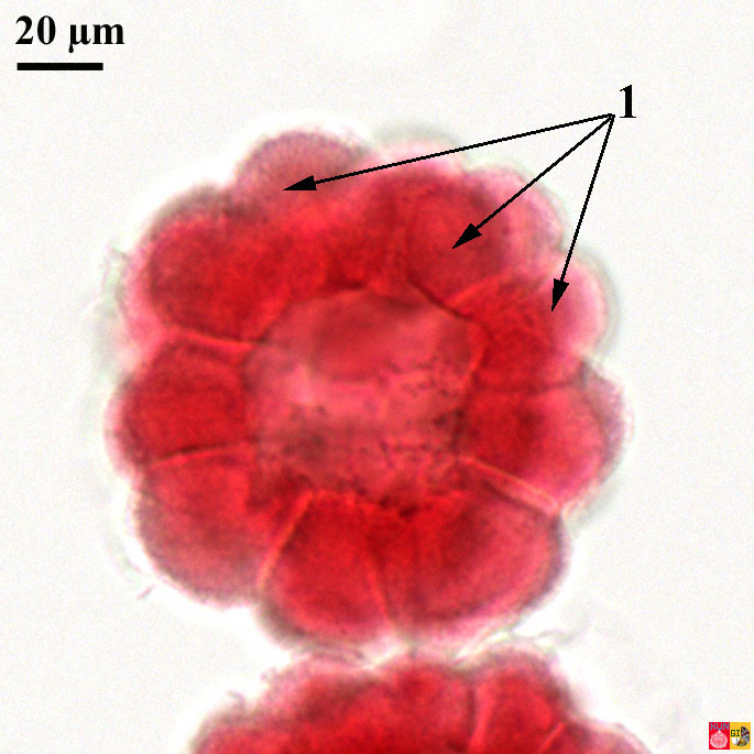
During the consecutive cleavage rounds respectively 2, 4, 8, 16, 32, 64, 128 and finally ends in the morula stage. After a while it becomes difficult to distinguish each single cell. Here the cluster probably consists of 32 cells.
Stained microscopical preparation of the 32 cells stage 1 Blastomeres
Morula stage
Information:
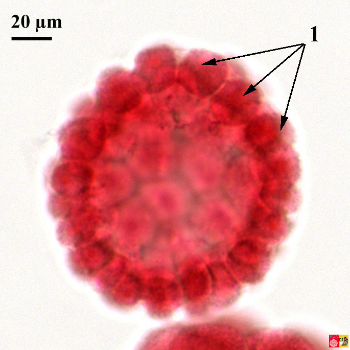
The pluricellular stage (here shown are probably 128 blastomeres) finishes in a morula (morula = mulberry). In this stage the cytoplasm of the fertilized egg is completed divided over the various blastomeres. Notice that this group of embryonic cells has about the same size as the original fertilized egg cell!
Stained microscopical preparation of the morula stage (about 128 cells).
1 Blastomeres
Blastula stage
Information:
After the morula stage the blastomeres spread and build a monolayer of cells around a central cavity (the blastocoel, bc). This stage is called the blastula stage (blastula = vesivle). The fertilization membrane is degraded by enzymes and disappears. The epithelium of the cells of the vegetal pole (vp) becomes thicker and forms the vegetal plate.
Stained microscopical preparation of the blastula stage 1 unequal blastomeres, 2 Blastocoel
Gastrula stage
Information:
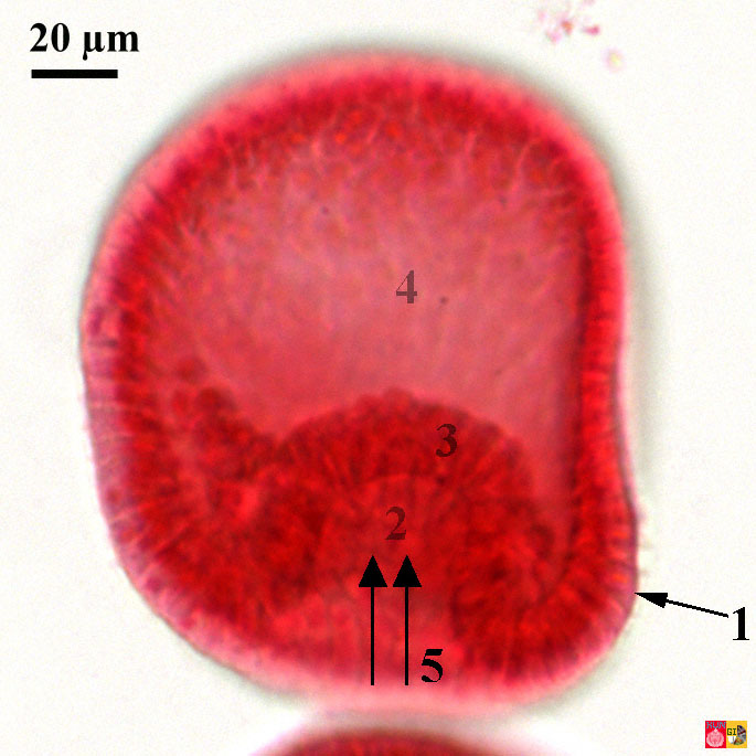
Blastomeres at the vegetal half migrate inward during the so-called invagination process (according tot the arrows in 5). In this way the ectoderm (ec) and the endoderm (en) are formed. The endoderm surrounds the developing primitive intestine or archenteron (arche = original-primitive, enteron = gut).
Stained microscopical preparation of the early gastrula stage 1 Ectoderm, 2 Beginning of the archenteron, 3 Endoderm, 4 Blastocoel (central), 5 Direction of invagination
Information:
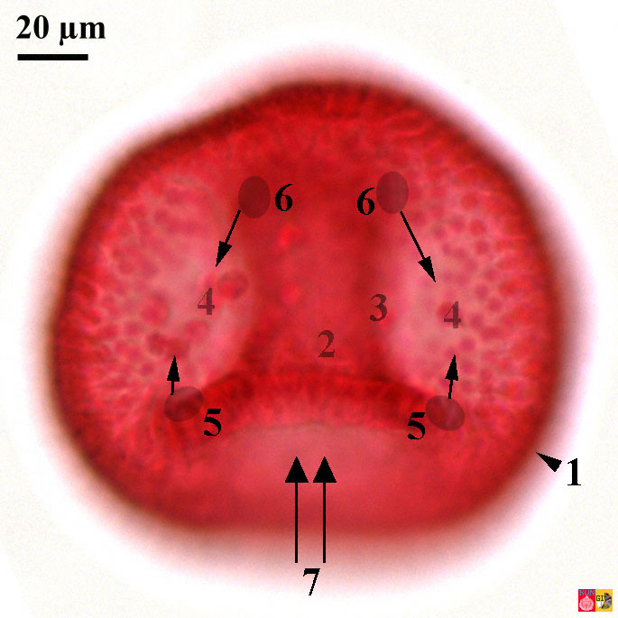
Shortly after invagination, individual cells at the base (5) of the growing archenteron (2) migrate to the inside (4). This process is known as the ingression of the primary mesenchym cells. This first group of migrating mesoderm cells (= mesenchym = mes) will later form the skeleton. A second group of mesoderm cells (6) separates from the upper part of the archenteron and forms the body cavities of the sea urchin.
Stained microscopical preparation of the gastrula stage (continued) 1 Ectoderm, 2 Archenteron (central), 3 Endoderm, 4 First group of mesoderm cells, 5 Origine of the first group of migrating mesoderm cells, 6 Origine of the second group of mesoderm cells, 7 Invagination area
Pluteus larval stage
Information:
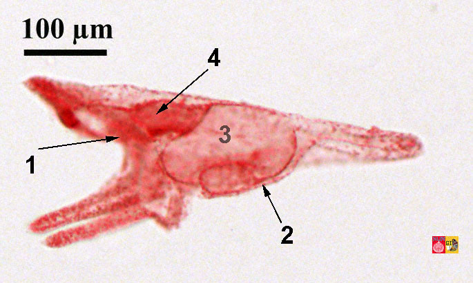
The final gastrula gives rises to the pluteus larva in the sea urchin. The Pluteus larva is primarily bilateral (= two-sided symmetrical). The radial symmetry in adult Echinodermata is a secondary phenomenon.
Stained microscopical slide of the pluteus (larva) 1 Mouth region, 2 Anus region, 3 Stomach (in transparence), 4 Intestine
Video of the moving parts of the developing gut: 2009 MBL Logan Science Journalism Program in Woods Hole
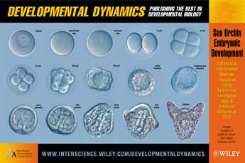
Overview of all stages
Free poster of Wiley.com about the embryology in the sea urchin
pdf; 440KB
Free poster of the Radboud University Nijmegen about the embryology of the sea urchin
jpg; 3000 x 4497 px; 2.1 MB