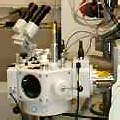
In order to be observed with a SEM, objects need first to be made conductive for current. This is done by coating them with an extremely thin layer (1.5 - 3.0 nm) of gold or gold-palladium. Furtheron, objects must be able to sustain the high vacuum and should not alter the vacuum, for example by losing water molecules or gasses. Metals, polymers and crystals are usually little problematic and keep their structure in the SEM. Biological material, however, requires a prefixation, e.g. with cold slush nitrogen (cryo-fixation) or with chemical compounds. This particular microscope is forseen of a special cryo-unit where frozen objects can be fractured and coated for direct observation in the FESEM (see pictures and more explanations at the
Cryo-SEM page). Chemically fixed material needs first to be washed and dried
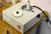
over the critical point to avoid damage of the fine structures due to surface tension. As an alternative to this tedious step chemical drying with hexamethyldisilazane is sometimes applied nowadays. After drying, coating with gold is performed in a separate device.

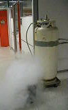
A lot of liquid nitrogen is consumed (container with liquid nitrogen in the picture here above left) to freeze material, maintain it cold in the cryo-unit,and to keep all pumps working. Hear the tunes of pouring liquid nitrogen in an isolating Dewar flask (a bit like a thermo flask) as
wav or
mp3 sound or see a short movieshot in
mov.
| Preparation of the SEM material |

|

|

|

|

|
| 1 |
2 |
3 |
4 |
5 |
Click on images for a zoom:
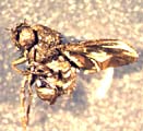
1 Pistil and drosophila coated with a thin layer of gold to permit good conductance of electrons in the SEM.
2 Nitrogen slush freezing unit.
3 Small container with liquid nitrogen.
4 Cryo-fracture coating unit |