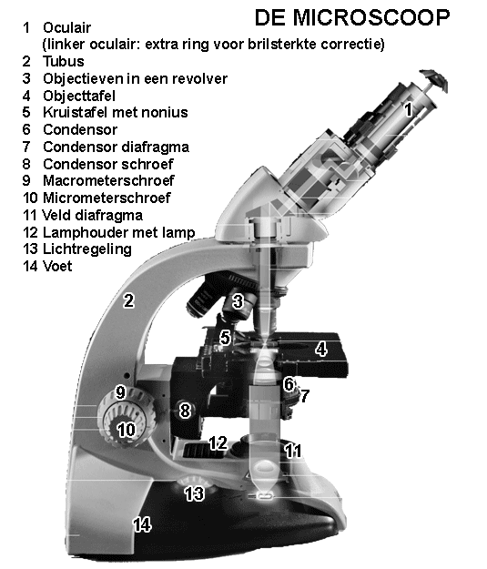
Study microscope |
||

THE PARTS OF TE MICROSCOPEThe English version of this webpage presents only a summary of the main items of the training microscopes used by the biology students at the Radboud University Nijmegen. However, the parts of the microscope described here as well as their function are common to almost all light microscopes.The student microscope has a heavy and strong bow-shaped standard (16) with a foot (15). All the parts hang on this frame, which is solid and stable enough to absorb most vibrations. The main five parts of the student light microscope are:
1. The light source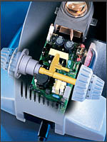 The light source consists of a halogene lamp that gives bright white light. The power cable can be connected to a normal 220V source. The intensity of the lamp (13) can be regulated with the illuminated knob (14). The light is concentrated by a simple collector lens. The light beam is deviated by a mirror through an opening in the frame to the object. By adjusting the opening of the field diaphragm (12) to the size of the field of view, stray light and disturbing scattering can be prevented, without infering on the brightness. The light source consists of a halogene lamp that gives bright white light. The power cable can be connected to a normal 220V source. The intensity of the lamp (13) can be regulated with the illuminated knob (14). The light is concentrated by a simple collector lens. The light beam is deviated by a mirror through an opening in the frame to the object. By adjusting the opening of the field diaphragm (12) to the size of the field of view, stray light and disturbing scattering can be prevented, without infering on the brightness.
2. A pair of lenses to bundle the light and project it on the objectThe light emaning from the field diaphragm (12) is focussed by the condensor (6; a system consisting of two lenses) on the objec. The focus point of the condensor must nbe adjust in such a way that the light is fully concentrated on the specimen, in the middle of the field of view, while illumination is even. The height odf the condensor can be adapted with knob 9, the centering of the light beam can be achieved with the two knobs 8 for perpendicular orientations. The condensor itself also bears a diaphragm, the condensor-diaphragm (7), that limits the light beam.3. The stage on which the object is positioned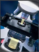 The microscope slide with the object can be clamped with a feather on the object stage (4). The slide holder is mounted on a mechanical stage (5) with axes that can shift in the X and Y-direction. There is a Vernier ruler (5; like on a caliper) that can be used to determine the exact position of an object and find it later back. The microscope slide with the object can be clamped with a feather on the object stage (4). The slide holder is mounted on a mechanical stage (5) with axes that can shift in the X and Y-direction. There is a Vernier ruler (5; like on a caliper) that can be used to determine the exact position of an object and find it later back.
4. A number of lenses for magnifying the objectAbove the stage there are 4 cilinders with compound lenses (3), the objectives (a 4x, 10x and 40x dry objective and a 100x oil immersion objective). The objectives are mounted on a rotating revolver (Lat. revolvere = rotate). The objective forms an enlarged image of -part- of the object. This image deviated by a mirror and projected through the tube onto the oculars (1) (Lat. oculus = eye) for a second enlargement. (The resolution depends however on the light source, the quality of the objective and the correct adjustment of the microscope in particular the level and opening of the condensor). One of the two oculars allows some correction for difference between the twoeyes and also the distance between the oculars can be adjusted to that between the two pupils.5. The focus knobs (10 and 11)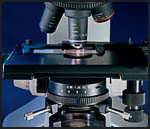 The distance between object and objective can be controlled with a coarse macro (10) and a fine micro (11) focus knob. The distance between object and objective can be controlled with a coarse macro (10) and a fine micro (11) focus knob. Links
|
||
