As help file for the understanding of the webpages on the cell cycle a number of terms and facts related to chromosomes and chromatids are highlighted here:
Chromosomes and chromatids occur as rod or thread-shaped structures in the nucleus of eukaryotic cells. Prokaryotes usually have only a single circular chromosome (in the webseries we will not focus on this group). The name chromosome (from the Greek chroma = color and soma = body) originates from the fact that chromosomes can be observed as stainable bodies in a light microscope during cell division.
Chromosomes and chromatids contain chromatin, which mainly consists of extremely long stands of DNA material (Deoxyribonucleic acid) that functions as carrier of genes and regulatory elements. Besides, chromatin contains Histones (chromosome proteins) and other proteins involved in the packaging of the DNA strands during condensation at cell division (see figure E here below) and small quantities of RNA.
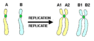
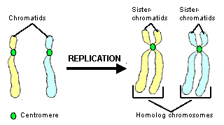
Sister chromatids (with -tid at the end), are two identical (= exactly the same) parts (Chromatids) arising from replication of a chromosome. (In the figure here next the sister chromatids A1 and A2 are an exact copy of each other, as well as the set B1 and B2, whereas homologs A's and B's show small differences). These two parts retain the denomination chromatids as long as they are bound together through the centromere, which is for example the case during the entire S phase following duplication of DNA (replication). This connection is vible as a constriction during mitosis or meiosis. During anaphase in mitosis and anaphase II in meiosis the two sister chroamtids are pulled apart at the centromer. According to current definitions, each single chromatid is regarded as a own chromosome after separation of the chromatids during cell division.
In most organisms chromosomes occur in pairs, the so-called homolog chromosomes (homolog = similar/corresponding). In contrast to the sister chromatides the homolog chromosomes consist of two (slightly) different copies of the same chromosome; homolog chromosomes carry yet the same genes, but the two copies of each allel can be either identicalor different of each other.
A single chromosome contains only on single long unbranched double-stranded DNA molecule that displays the typical double-helix structure. This double-strands DNA is formed by one phosphate group alternating with one desoxyribose group coupled to each other by nucleic acids (adenine, guanine, thymine en cytosine). These nucleic acids form consistent pairs (AT and CG). Of each complementary strands of the DNA molecule the antisense can be read (used as a template) for the synthesis of proteins, but not the sense.
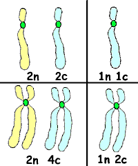
The ploidy refers to the number of different copies of each chromosome present in a cell. Most plants and animals are diploid, indicated by 2n, which means that there are twocopies of each chromosome per cell. Their gametes, however, are haploid, indicated by n (one single copy). Bacteria and some plants and fungi are haploid organisms.
The number of chromatids or chromosomes coding for the same (corresponding) genes within a cell, is sometimes indicated by the small letter c. For example, a cell that was 2c before replication, will become 4 c after replication, thus when the DNA has been doubled in preparation of mitosis, because four samples of DNA stands coding for the same genes are present, but the ploidy will remain unchanged: if the cell was 2n, it is still 2n after replication and it was 4n it remains 4n.
Chromosomes,chromatids, centromeres and telomeres
2n 2c means two homolog (diploid) unreplicated chromosomes (two chromatids). 1n 1c one single chromosome (haploid) that is unreplicated. 2n 4c Two homolog chromosomes (diploid) consisting each of two sister chromatids (two yellow and two blue), thus 4c in total. 1n 2c one single chromosome in which DNA has been duplicated.
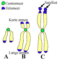
A. Telocentric, B. Acrocentric en C. Metacentric chromosome. Chromosomes are classified on account of their size, location with respect to the centromere and presence or absence of satellites
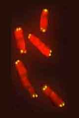
D. Computer-generated illustration based on a microscopic photograph: red = chromosomes, yellow fluorescence = telomeres labeled with a specific probe
Besides hosting the genes that function as archive for genetic information, chromosomes also bear pieces of DNA between the genes which have a structural function. This is the case for the telomere and centromere that are involved in replication and cell division.
The centromere (centron = middle, meros = part) is the region of the chromosome where the chromatids that arise from replication are held together. The centromere hosts the kinetochore, a protein complex where the spindle filaments attach during mitosis or meiosis. Because the centromere remains relatively little spiralized during prophase and metaphase it can be distinguished as a primiry "pinch".
The telomere (Gr. telos = end, meros = part) is a piece of DNA that is located at the end of the chromosomes and serves to prevent DNA corruption. Strongly repeating, so-called satellite sequences are related to telomeres. During replication sometimes part of the telomere are lost so that they appear shorter after cell division. When the enzyme telomerase is present in the cell telomeres can recover their length, but too severe shortening leads to chromosome instability and irreversible damage so that divison is impeded and cell death eventally occurs. On the other side, cells with active telomerase can continue to divide.
Structuur van chromosomen
Klik op figuur voor een Zoom. (Folded chromosomes are clearly visible under a light microscope)
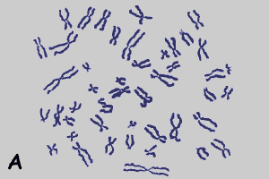
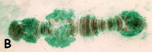
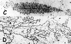
Figure A and E: Computer-generated illustrations; B-D: micrographs (J. Derksen).
A Karyogram of a human (Homo sapiens lymphocyte. As a standard procedure cells are stained with Giemsa's and observed with a light microscope at the metaphase stage. In that phase the two chromatids of the chromosomes can be easily discerned. Chromosome duplication has been accomplished in the previous S-fase.
B Giant chromosome from the salivary gland of the midge Chironomus tentans, consisting of many chromatids produced by rounds of endomitosis following synapsis of the two homologues. The chromatin has been stained with Orcein. The darkly stained areas are regions with condensed chromatin (bands), the lighter stained areas are regions with less condensed chromatin (interbands). The large bulges or Balbiani rings are regions with a very high RNA synthesis. These have been stained with Fast Green here.
C A ribosomal gene from the nucleolus of Drosophila (the fruitfly). (Ribosomal genes produce the ribosomes consisting of subunits made up of proteins and rRNA, which control the translation of messenger RNA into proteins.) In this typical "Christmas tree structure" the gene occupies the place of the "trunk" and RNA/protein side-chains increase in length while synthesis proceeds. The preparation has been made by dispersion of the chromatin in a basic solution followed by imaging in an electron microscope (" Miller spread").
D Image of a gene coding for a protein. This gene produces messenger RNA. The image was made in the same way as in C. In comparison to C, the side chains of this protein-encoding gene are less numerous but longer and they are often folded.
E Schematic representation of the packaging process from a DNA molecule to a condensed mitotic chromosome.
Part 1 of this animations illustrates the folding of DNA into a chromosome. Source: You Tube; Credits to the authors
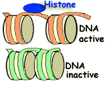
A chromosome consists of a long DNA molecule that is folded around proteins. Especially the histones (positively charged proteins with arginine and lysine-rich, strongly alkalic regions) play a role in "packing" the DNA, because they bind to the negatively charged DNA molecule. DNA and chromosome proteins together are also called chromatin.
During normal cell activities only a selection of the DNA in the chromosomes is unfolded. In domains where the DNA codon is read out, the molecule is even completely open to give access to enzymes. However, at at the beginning of cell divisions, in both mitosis and meiosis, the DNA gets strongly spiralized, a configuration called condensation or spiralisation. Chromosomes can be more conveniently separated in this compact form. (Figure E).
Number of chromosomes and chromatids
The number of chromosomes is indicated by a small letter n.
Normal body cells of diploid organisms always contain pairs of homolog chromosomes. All these cells are in principe genetically identical to the zygote (the product resulting from fertilization) from which they descend. Each pair of homolog chromosome is a heritage of one set from mother side and another from father side. However, because of meiotic "reshuffling" (= crossing-overs), a different pool of genes is present in the inherited chromosomes than in the cells of the father and the mother!
Sexual reproductive cells have only one sample of each chromosome.
An unreplicated chromosome contains one double strand -DNA molecule.
A replicated chromosome contains two identical double strand -DNA- molecules, the chromatids, that are joined at their centromere.
Two single sets of pairs (daughter) chromosomes are formed by separation of the pair of chromatids during cell division (from anaphase on in mitosis and anaphase II in meiosis)
Haploid is the term for a cell or an organism with only one set of chromosomes (n). If this is valid for all cells, the entire organism is regarded as being haploid. A haploïde cell remains in fact n after replication, but doubles from c to 2c. Each chromosome consists of two chromatids.
Diploid is the term for cells with a double number of chromosomes (2n), whereby one set of chromosomes is homolog to the other. (The sex chromosomes present in each human cell are an exception). A diploid cell remains 2n after replication, but doubles from 2c to 4c.
Humans usually have 22 pairs of homolog chromosomes (autosomes) and as sex chromosomes 2 x chromosomes (in female), or 1 x and 1 y chromosome (in males), thus in total 46 chromosomes per body cell (see human karyogram; 2n = 46) and 92 chromatids per cell after replication.
 A chromosome consists of a long DNA molecule that is folded around proteins. Especially the histones (positively charged proteins with arginine and lysine-rich, strongly alkalic regions) play a role in "packing" the DNA, because they bind to the negatively charged DNA molecule. DNA and chromosome proteins together are also called chromatin.