Amphibians: in sections |
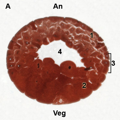
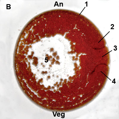
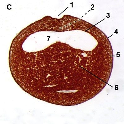
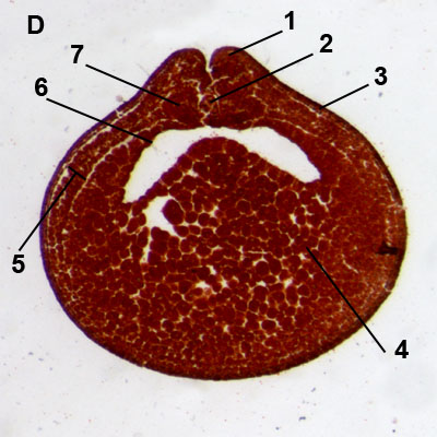
This page presents micrographs and explanations on a number of characteristic stages of the embryonic development in the common frog (formerly Rana esculenta, nowadays Pelophylax sp.) and the tadpole ( Xenopus laevis) viewed in stained cross sections.
| Early developmental stages in the common frog) sections) |
A. Blastula
An = Animal pole, Veg = Vegetal pole, 1 = Micromeres, 2 = Macromeres, 3 = Marginal zone, 4 = Blastocoel
|
B. Gastrulation: yolk plug stage
An = Animal pole, Veg = Vegetal pole, 1 = Ectoderm, 2 = Dorsal blastoporus lip, 3 = Yolk plug in blastoporus, 4 = Ventral blastoporus lip, 5 = Remnants of blastocoel
|
 |
 |
C. Neurulation: neural plate
1 = Neural plate (just before the folding), 2 = Area of chorda mesoderm, 3 = Endoderm, 4 = Ectoderm, 5 = Mesoderm, 6 = Yolk material, 7 = Archenteron |
D. Neurula stage: neural groove
1 = Neural fold, 2 = Chorda, 3 = Ectoderm, 4 = Yolk material, 5 = Lateral plate mesoderm, 6 = Endoderm, 7 = Dorsal mesoderm (somites)
|
 |
 |
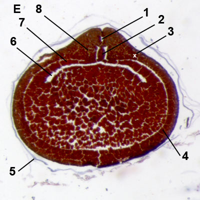
E. Neurula stage: neural tube
1 = Neural tube, 2 = Chorda, 3 = Area of intermediate mesoderm, 4 = Lateral plate mesoderm, 5 = Remnants of fertilization membrane, 6 = Archenteron, 7 = Endoderm, 8 = Somite mesoderm | |
 |
|
| Larval developmental stages in the tadpole (sections) |
It is recommended to study these images along with the textbook:
Atlas of descriptive embryology. Gary C. Schoenwolf. Pearson, Benjamin Cummings eds. ISBN 978-0-13-158560-7 |
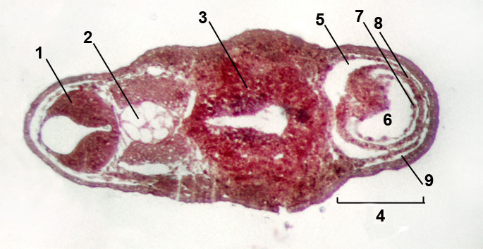 |
Larval stage: heart region:
Ref. figure in textbook (7th edition): 6.36
1 = Rhombencephalon, 2 = Chorda, 3 = Intestine, 4 = Heart region, 5 = Pericardial cavity, 6 = Heart tube, 7 = Endocardum, 8 = Myocardum, 9 = Fixation artifacts |
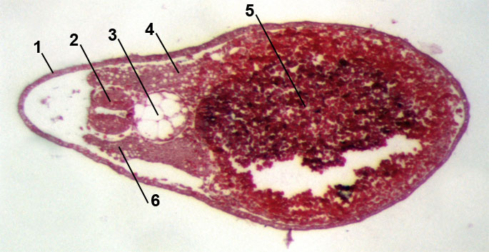 |
Larval stage: tail region:
Ref. figure in textbook (7th edition): 6.38
1 = Ectoderm (epidermal), 2 = Neural tube, 3 = Chorda, 4 = Mesoderm, 5 = Endoderm cells filled with yolk, 6 = Somite |
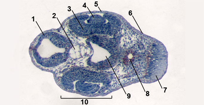 |
Larval stage: head region / formation of the eye:
Ref. figure in textbook (7th edition): 6.47 and 6.48
1 = Neural tube (detail rhombencephalon), 2 = Chorda, 3 = Retina, 4 = Lens, 5 = Cornea, 6 = Ectoderm (epidermis), 7 = "Adhesive gland", 8 = Foregut, 9 = Diencephalon, 10 = Developing eye |
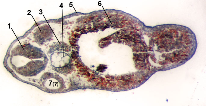 |
Larval stage:
Ref. figure in textbook (7th edition): 6.49 and 6.52 (here sectioned obliquely in L-R profile)
1 = Neural tube (spine), 2 = (Somite) derma myotome, 3 = (Somitet)sclerotome, 4 = Chorda, 5 = Ectoderm (epidermal), 6 = Yolk, 7 = (probably) Ear vesicle |
|
|


