Chicken: 18-20 h |
Using drawings and micrographs of stained samples the early embryonic development in the chicken is depicted. The images of the in toto (whole mount) preparation show processes in the geminal disc (also called blasto disc) viewed from above, as if one would observe the embryon at the yolk surface through a window in the hatching egg. Also photographs and schemes of cross sections are available.
Early embryonic developmental features that are shown on this page:
Earliest developments in the germinal disc (More...)
Whole mount preparation 18 hours (More...)
Whole mount preparation 20 hours (More...)
Earliest development phases of the germ layer
The embryonic development in birds occurs in the germinal layer separated from the upper side of the yolk by the subgerminal cavity. In the early stages, the germinal layer confines on the yolk with the area opaca (opaca = untransparent) that appears as a dark zone at the external border of the germ layer. The central region of the germ layer is localized at the upper part of a hole (the blastocoel). This region is transparent and is therefore called the area pellucida. The area pellucida is composed by two layers, the epiblast at the surface and the hypoblast in the deeper part. The gastrulation process starts with the formation of a groove and forms first the primitive groove at the caudal side. As this groove looks as a streak, it is also called the primitive streak. The cells in the central zone divide frequently and migrate in laterally between the epiblast and hypoblast.
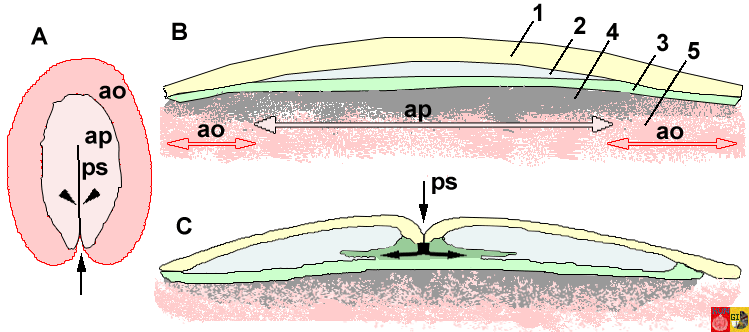
In a early stage, the area opaca appears as a dark zone at the external border of the germ layer and is in contact with the yolk mass. (opaca means untransparant; see for example in fig.A). The central region of the germ layer is localized at the upper part of a hole (the blastocoel). This region is transparent and is therefore called the area pellucida (ap in A). The area pellucida is composed by 2 layers, the epiblast (1 in B) at the surface and the hypoblast (3 in B) in the deeper part. The gastrulation process starts with the formation of a groove (see arrow in fig C, transversal section) and forms first the primitive groove at the caudal (tail?) side. As this groove looks as a streak, it is also called the primitive streak. The cells in the central zone divide frequently and migrate in laterally between the epiblast and hypoblast (see lateral arrows in C).
| Schematic representation of parts of the germinal disc during early embryonic development |
 |
The germinal disc in upperview (A), and cross section before (B) and during (C) gastrulation.
ap = area pellucida, ao = area opaca, ps = primitive streak, 1 = Epiblast (forms the ectoderm), 2 = Blastocoel, 3 = Hypoblast (forms the endoderm), 4 = Subgerminal cavity, 5 = Yolk |
Stage 18 hours
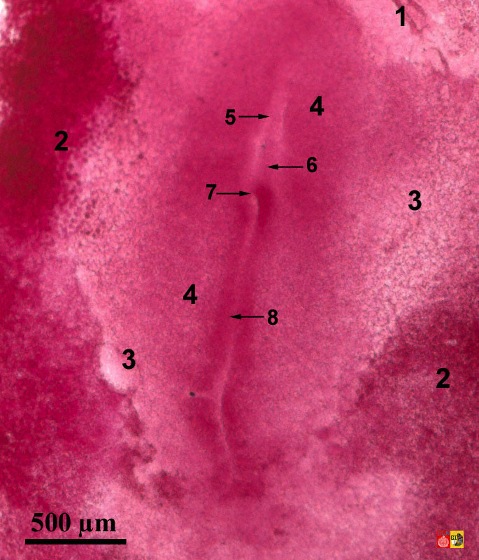
At 18 hours after fertilization, the area pellucida (= transparent) is surrounded by the dark area opaca (= not transparent). The primitive streak lies in the middle of the area pellucida as a clear line. At the tail side of the area pellucida it is characterized by a primitive groove surrounded by 2 thicker walls of the blastoderm, the primitive walls. In the front, the primitive streak ends in a small pit, the primitive pit or Hensen's node. This structure is homologue with the dorsal lip of the blastopore in amphibians. Just in front of this node one can distinguish the formation of the notochord (chorda). This structure induces the formation of the neural plate and later the neural groove. In the embryo stage around 20 hours after fertilization, the head fold is visible as a half circle that lifts up from the subcephalic pocket in the front of the germ layer. The proamnion lies in front of (= pro) this head fold as a clear zone. The amnion is one of the membranes that will protect the embryo in a later stage. This stage is characterized by 3 pairs of somites. The neural groove becomes visible and blood islands are formed in the area vasculosa which is the inner part of the area opaca. The peripheral region of the area opaca, the area vitellina is not vascularized.
| Wholemount preparation 18 hours |
Information:
In the cenrtal region, you see the area pellucida (= transparent) surrounded by the dark area opaca (= untransparant). The primitive streak lies in the middle of the area pellucida as a clear line. At the tail side of the area pellucida it is characterized by a primitive groove surrounded by 2 thicker walls of the blastoderm, the primitive walls. In the front, the primitive streak ends in a small pit, the primitive pit or Hensen's node. Just in front of this node one can distinguish the formation of the notochord (chorda). This structure induces the formation of the neural plate and later the neural groove. |
 |
Embryology of chicken 18 hours after fertilization: stained whole mount preparation
1 = Proamnion, 2 = Area opaca (dark), 3 = Area pellucida (transparent), 4 = Embryonal region, 5 = Neural plate, 6 = Chorda, 7 = Hensen's node, 8 = Primitive streak
|
Stage 20 hours
| Whole mount preparation 20 hours |
Information:
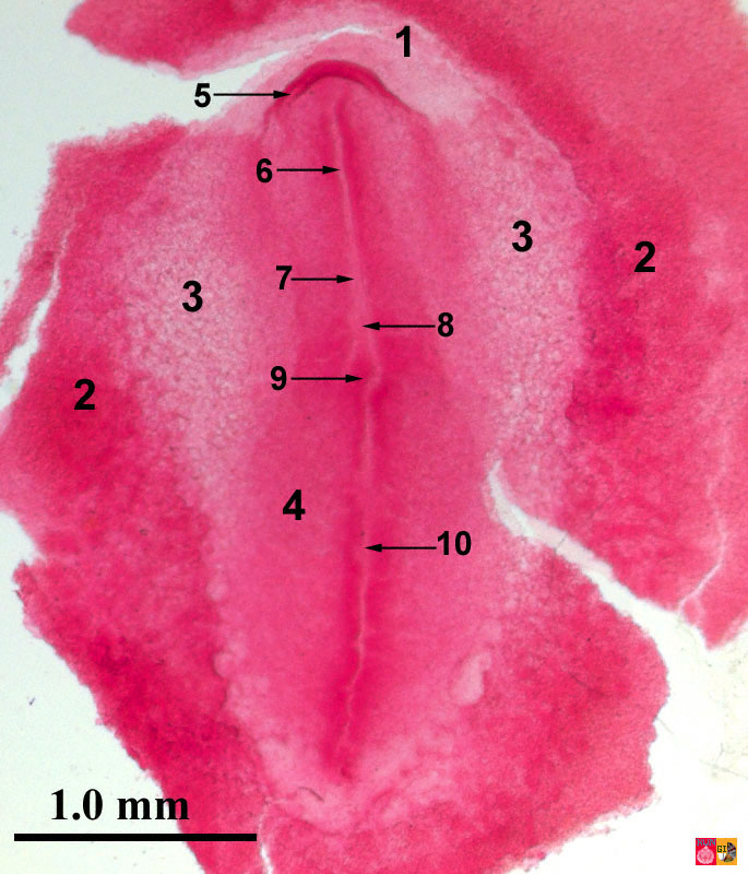
In the embryo stage around 20 hours after fertilization, the head fold is visible as a half circular elevated structure in the front of the germ layer. The proamnion lies in front of (=pro) this head fold as a clear zone. The amnion is one of the membranes that will protect the embryo in a later stage. |  |
Embryology of chicken 20 hours after fertilization: stained whole mount preparations
1 = Proamnion, 2 = Area opaca (dark), 3 = Area pellucida (transparent), 4 = Embryonal region, 5 = Head fold, 6 = Neural groove, 7 = Neural plate, 8 = Chorda, 9 = Hensen's node, 10 = Primitive streak
|
|
|


