|
 |
Examples of cryo-SEM views:
left entire object (here seedling), right freeze-fractured sample (here leaf)
Imaging: M. Wolters-Arts, GJ Janssen and W. Kerkhof |
| Click on the magnifier and drag the cursor PRESSED DOWN to the image hereabove and release the cursor. A zoom window appears. This magnifier can then be moved over the entire image. To (re)reactivate the magnifying function, refresh the browser (press shortcut key F5 in IE). Magnifier javascript: http://valid.tjp.hu/zoom |
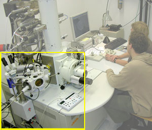
In a cryo scanning electronen microscope (cryo-SEM; cryo =cold) images can be made of the surface of frozen material, i.e. entire cells and small pieces of tissue (e.g. here left a seedling of Arabidopsis from the third-year practical course on "Hormones in Plant Development"), but also of fracture surfaces inside the original specimen (here left, cleaved leaf of elderberry in which epidermis, palisade parenchyma, sponge parenchyma and a vascular bundle are visible).
The cryo-method has the great advantage that the material is so rapidly frozen that vulnarable biological structures are well preserved and that when using cryo-fracture the material breaks clean along weak edges (e.g. membranes) without causing much malformation.
Microscope and preparation of cryo-samples
The material is fixed on a holder with a layer of carbon-rich conductive
glue (conductive to allow discharge of electrons). It is rapidly frozen with -boiling- liquid nitrogen (
mov or
gif movie of the sample and its holder in boiling nitrogen) or
'nitrogen slush' that transmits cold well. The holder with frozen material is held under liquid nitrogen to be coupled to a
rod and pulled back into a small
cylindrical container. This is done to transfer the sample to the high vacuum cryo-unit and prevent too much contamination with gas particles while
sliding in the sample into the cryo-chamber. The cryo-chamber is equipped with a knife that can be handled from outside by means of a level to fracture the sample for applications in which imaging of the surface of innerstructures is aimed. Next, disturbing humidity from the sample is sublimated and a thin layer of gold-palladium is "sputtered" on the material, this again for good conductance of electrons. Finally the entire or fractioned material is further inserted into the observation chamber with a rod.
| Cryo-SEM: front side and rear side! |
 |  |
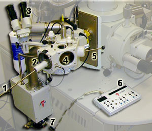
Parts of the cryo-unit in the FESEM:
1 = insertion rod, 2 = cylindrical chamber for transfer of the sample from freezing unit to cryo chamber, 3 = binocular, 4 = cryo-chamber proper containing breaking knife, gold pallladium sputter, water decontamination sublimation unit, 5 = lever to handle the fracture knife, 6 = operation display, 7 = supply of liquid nitrogen to the cryo-FESEM
|
| Cryo-SEM: preparative manipulations |
 |  |
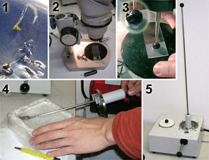
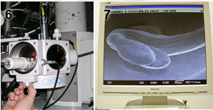
Processing:
zoom
1 = sample (here seedling), 2 and 3 = fixing the sample to the holder using a carbon-rich conductive glue, 4 = freezing of the sample in liquid nitrogen (or nitrogen slush, not shown on this picture), 5 = transfer of the sample to a closed cylindrical container , 6 = insertion of the sample into the cryo-chamber, 7 = resulting image on the monitor |
Wikipedia page on
electron microscopy 


