What does the word FESEM mean?
FESEM is the abbreviation of the word Field Emission Scanning Electron Microscope. Scanning and electron transmission microscopes (SEM and TEM) use as a source for image formation electrons (particles with a negative charge), in contrast to light microscopes (LM). These electrons are produced by a Field emission source in a FESEM. The sample (object) is scanned in a kind of zig-zag pattern by an electron beam.
 Zoom in
 on a diagram of the beam pattern in a light microscope (LM), a transmission electron microscope (TEM) and a scanning electron microscope (SEM) (The original black & white drawings are from Jeol. Used with permission).
What can be done with a FESEM?
A FESEM is used to visualize very small topographic details on the surface or entire or fractioned objects. Researchers in biology, chemistry and physics apply this technique to observe structures that may be as small as 1 nanometer (= billion of a millimeter). The FESEM may be employed for example to study cell organelles and DNA material, synthetical polymeres, and coatings on microchips. The microscope that has served as an example for the virtual FESEM is a
Jeol 6330 that is coupled to a special freeze-fracturing device (Oxford Ato). The real FESEM is located at the Department of
General Instrumentation of the Radboud University Nijmegen.
How does a FESEM function?
Electrons are liberated from a field emission source and accelerated in a high electrical field gradient. Within the high vacuum column these so-called primary electrons are focussed and deflected by electronic lenses to produce a narrow scan beam that bombards the object. As a result the "electron hail" secondary electrons are dislocated from each spot on the object. The smaller the angle of incidence of the electron beam is with respect to the sample surface and the higher a certain point is in the sample, the more secondary electrons are able to reach the detector and the lighter this dot will appear in the final image following electronic signal amplification and digitalization. Also the composition of the sample has an effect on the number of deflected electrons too and thus on the 'gray value' of the corresponding pixel in tthe final image. (Of course, the position of each point in the sample relative to the detector is of great influence on the strength of the signal, but this distance factor is taken into account and compensated). Detection of the secondary electrons results in a kind of
three-dimensional shadow-cased surface representation of the sample.
To prevent that electrons that are not captured by the detector would hang like a cloud masking around the sample, thus masking the image, SEM samples are coated in advance by a very thin layer of conductive material, e.g. gold-palladium or carbon or platinum, to clear away superfluous electrons.
How does een cryo-FESEM look like?

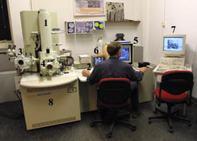
A cryo-FESEM looks like a large desk on which a cylindrical column is mounted, and behind which pumps and tubes can be found. The column (1) hosts in the upper part the electron gun, and at various levels below electro-magnetic lenses to align the electron beam. Tubes and pumps are involved in maintaining the vacuum inside the instrument and these are powerful cooling units to keep the temperature in the cryo-unit far below zero. The microscope is operated from the steering panel (2; on the desk). A close copy of this panel has been used for the simulations. The cryo-unit with a binocular (3) is located left of the column. When conventional (not cryo) microscopy is applied the exchange chamber in front, below the columns (4) is used to introduce the object into the high vacuum area. The object can be observed on the large screen (5) while it is scanned. The small screen (6) serves to watch the object chamber. The computer for image archiving and processing is located right (7). The cupboards below the desk contain (LOT OF) electronics (8). On the background the sound (
wav or
mp3 sound) of the
pumps that maintain the vacuum in the column can be heared as well as the sissing of the boiling nitrogen for the freeze-unit and the cooling of the column (sound when tapping liquid nitrogen into a container in
wav (388KB) or
mp3 (87KB)).
Wikipedia page on electron microscopy


