| Anaglyph animation with depth impression[ |
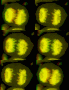 |
| Microscopical red-green anaglyph images corresponding to various projection angles have been mounted in a film sequence. From this series of images with depth information it can be determined that the daughter chromatides were already separated in this deviding cell (anaphase stage) |
Man has the very special ability to physically perceive depth and to be able to interprete three-dimensional shapes and movements in a spatial environment. This is because our two eyes are positioned next to each other. The images preceived by the retina of our left and right eye show just some shift and a slight rotation with respect to each other (
retinal disparity). This phenomenon is called
parallax. In the visual cortex of the brain these two mainly overlapping, but still slightly different stereo images are combined to one whole. A sensation of depth vision arises and understanding of the spatial arrangement around us, which is quite fascinating (more about
binocular vision = vision with two eyes). Our field of vision remains relatively narrow and one can not speak of true 3D vision. When we watch an object we only see the side directed towards us and a little bit of the side flanks, but the rear side remain hidden for us. Except for some teachers, man can also not see what happens in his/her back. We also lack eyes on stalks which would enable us to scan all facets of an object and we are also not equipped with a ten-eyes system like a Google streetview camera! According to the "
Optometrics network" at least 12% of people have some type of problem with their binocular vision. Remarkedly, with only one eye it is often still possible to make a good estimation of the spatial distribution of objects around us (this is also possible with other senses, by the way!): we can scan the surrounding by directing our eyes and head, while our eyes stepless focus on the shapes on which we concentrate. From this bulk of visual information coupled to earlier experiences, in particular distortions related to perspective, the size and significance of objects, we are able to re-create a "depth picture" in our head.
| Depth vision |
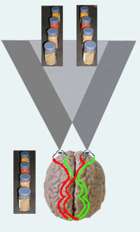 |
Depth perception: phenomenon of parallax: our two eyes perceive two slight different windows of the world (see schematic representation).
Test: put a few objects (here jars with spices) at a distance of about 1 meter. Keep the head still and look alternately with the right and left eye at those objects (quickly alternate left and right): the objects (their view) seem to jump aside. When looking again with both eyes the two images appear in our impression to be automatically reconstitued to one quiet single unit. |  |
| Interpretation of depth from perspective and spatial context: in one wink it is possible to make up from this photograph which tree was the closest to the photographer and which ones were further away, even though the projection on the screen is in fact a flat image. From earlier experience, e.g. knowing the size of a swan, we can guess that the trees were big. |
Depth vision by means of visual tools provide a virtual or remote, sometimes magnified, stereo representation of the reality, i.e. stereo-binoculars, a stereocamera, -microscope, or -laparoscope, is not only fun (e.g. for games), and often spectacular (e.g 3D images of the
exploration of Mars, it can be truly useful, for example for accurate handling during surgery, for precise remote navigation of a space craft, or for a better understanding of the organization of a macroscopical (e.g. in
earth sciences) or microscopical object (find elwehere on this website a
microscopy stereo viewer).
Examples from microscopy
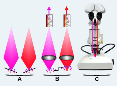
Three approaches to acquire depth information in a microscopical object are depicted here on a diagram of a dissection microscope, namely
A to tilt the object,
B to make series of images at various focus levels, or
C to keep the object in fixed position and take pictures at at least two angles.
| Acquisition of depth information and parallel stereo projections in microscopy |
 |
 |
|
Microsocpical object: horse statue above the Reichstag on a German 10 euro cent coin. |
 |
Ways to acquire depth information:
A One can physically tilt the object like in a seesaw (teeter-totter) while maintaining the center of the object in the middle axis of the microscope. The tilt should be done for at least two positions, usually a few degrees in positive and negative sense. Separate photographs are made in each position. This approach is widely used, in particular in Scanning electron microscopy (EM) and especially in Transmission EM tomography.
B. In confocal laser scanning microscopy it is common to make reflection or fluorescence and sometimes transmission images that correspond to thin slices in the object (optical sections) in het object, at increasing focus level. From such a series of xyz micrographs, comparable to a stack of pancakes, two with respect to each other slightly rotated projection can be produced with an imaging program that mimic , as if seen in depth vision through the left and right eye (see examples here below).
C One can also record the same object from on photograph or film under -at least two- different angles (about +3 and -3 degree with respect to the axis). This is one of the new trends in stereo and dissection-microscopes). |
Parallel projection
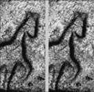
Probably the oldest method to display stereo is that in which two perspective-images corresponding to a view as seen from respectively the right and left eye, are print aside as photographs or projected next to each other on a screen (this method has been already applied in the pioneering years of electron microscopy). When having digital z-stacks of xy-images, like is often the case in confocal microscopy, these can been reconstructed to a duo-image that mimic two angles of view. Click to zoom in the example: the depth effect is best visible by gazing at about 50 cm distance from the monitor and while looking allow the two images to shift over each other and fuse to one view. In practice this method to view depth is for many people difficult; anaglyph projection, shown here below, works usually easier. |
3d rendering methods
In the course of time
rendering methods with static images or with animations have been developped to display the 3D structure of objects, among which microscopical samples, for example by means of:
- Parallel projections (see explanations here above and example)
- Color anaglyphs and polarized dual-images (see explanations here below and various anaglyphs, among which this example)
- Orthogonal projections (= projections along the perpendicular xyz-axes as here below; example)
- Shadow-cast projections (artificial representation as if light would shine on the object and elevate parts would cause shadow; example)
- Color-coded depth representation(= the color gives an indication of the depth in the object; see here below and example)
- Holograms and stereograms
- Various skin and relief representations (example)
| Orthogonal, shadow, color-coded and skin depth displays |
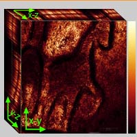 |
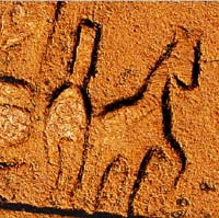 |
| Orthogonal 3D projection | Shadow-cast depth projection |
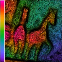 |
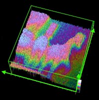 |
| Color-code (look-up table = LUT) depth-representation | Skin and wireframe rendering |
A few links on stereo vision
Software to make anaglyphs


