By combining light microscopy with image enhancement dynamic processes, like organelle movement, can be visualized within
living cells.
During the
video microscopy course that
Prof. dr. I.K.
Lichtscheidl
(University of Vienna) gave in the framework of a European collaboration program with the RU Nijmegen, special video and light microscopy techniques were demonstrated on tip-growing plant cells (here:
Medicago sativa root hairs and
Amaryllis pollen tubes).
The film sequences shown below are -compressed- real-time views acquired by employing high numerical aperture optics and monochromatic light (a.o. ultraviolet light with quarts optic), and by applying background correction and analog cq digital contrast enhancement.
| Technique and object
| Example
view
| Download film |
Bright field without background subtraction
Left: still image.
Movie right: germinated pollen grains in which the spindle-shaped generative
cell can be discerned, followed by a zoomed view of the movement of organelles
in the pollen tube (diameter about 10 micrometer)
|
 |
46s; real-time
wmv (1137 kb)
mpg (4053 kb) |
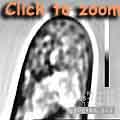
Video-Enhanced-Contrast (VEC)-bright field
By applying background
subtraction in VEC microscopy in conjunction with analog and digital contrast
enhancement, the presence and movement of small particles can be determined in
more detail, like here orgnalles in the tip of a root hair. Movie right: Chaotic
movement is typical for the extreme tip region, whereas directed movement can be
seen away from the apex
|
 |
42s
wmv (1211 kb)
mpg (3780kb) |
Phase-contrast
Phase-contrast does not only produce contrasted
images but also a kind of halo around particles. With this technique it is
therefore difficult to identify single organelles in the dense cytoplasm at this
pollen tube tip, but the technique works beautifully to image a thin layer of
cytoplasm (i.e. a vacuolate cell region) or bacteria
|
 |
38s; real-time:
wmv (1066 kb)
mpg (3373
kb) |
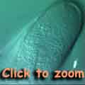
Differential Interference Contrast (DIC)
Particles and objects
differing in optical density appear with a shadow-effect. Thanks to this
property single organelles (for example mitochondria) can be tracked in living
cells, like in this pollen tube
|
 |
32s; real-time
wmv (803 kb)
mpg (2866 kb) |
Dark field
Dark field microscopy is an ideal technique to image
tiny particles (e.g. gold-particles for labeling, bacteria, irregularities in
the cell wall, silver grains in in situ hybridization, single
organelles). In this pollen tube organelles appear as bright objects in a dark
background
|
 |
32s; real-time:
wmv (843 kb) |
Ultraviolet
These views were made with UV light (370, 410, 440nm)
that is not suitable for direct eye vision, but can be well detected by a
special video camera. With this set-up very small particles, probably Golgi
vesicles (about 300 nm in size) that exhibit an extremely rapid chaotic
movement, can be observed along the cell membrane in de tip of this pollen
tube
|
 |
11s; real-time:
mpg (1600 kb) |
Colophone
Recommended literature as
rtf text or
PDF document
These images and movie sequences are
protected by the copyright terms of the Radboud Universiteit Nijmegen; in
contrast to other vcbio material these views are "intellectual" property of the
IECB 
Prof. Dr. Irene Lichtscheidl
Cell Physiology and Scientific Film
"IECB =
Institute of Ecology and Conservation Biology
University of Vienna (Austria)


