Structure of the DNA molecule

A
DNA molecule (deoxyribonucleine acid) is made of
two chains of several thousands of linked
nucleotides. Each nucleotide consists of a
phosphate group, a
deoxyribose and a
nitrogenous base (Fig. 1). The four
nitrogenous bases that occur in DNA are:
adenine (A),
guanine (G),
thymine (T) and
cytosine (C) (Fig. 2). Within each DNA strand, single
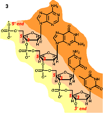
nucleotide-monomeres are bounded through their phosphate group to the sugar group of next monomere (Fig. 3).
| Structure of a DNA nolecule |
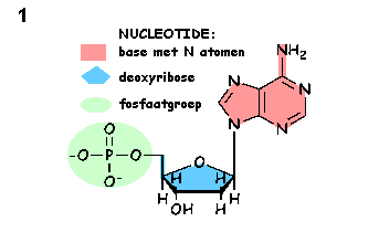
Fig.1. Structure of a nucleotide. Each nucleotide consists of a phosphate group, a pentose (= a sugar with 5 carbon atoms; in DNA deoxyribose) and a base containing nitrogen (in DNA: adenine, thymine, guanine and cytosine, in RNA adenine, guanine, cytosine and uracil instead of thymine).
|  |
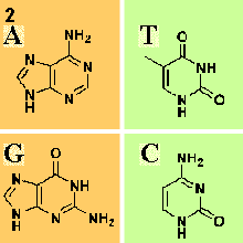 |
Fig.2. Structure of the nitrogenous bases from DNA. Adenine (A) and guanine (G) belong to the purines, while thymine (T) and cytosine (C) are both pyrimidines
|
|
Fig. 3. Bounds betwen nucleotide monomers in DNA.
Yellow = phosphate groups
Light-orange = sugar groups
Orange= nitrogenous bases
|
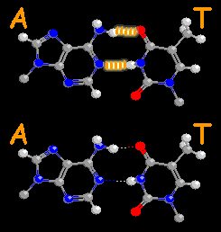
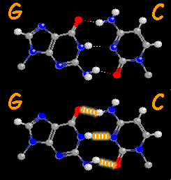
The two chains of the DNA molecule are linked together through hydrogen bridges between two opposite nitrogenous bases,
base pairs. each N-base has its own binding partner: adenine always with thymine (
A-T; Fig. 4) and cytosine with guanine (
G-C; Fig. 5).
| Base pairs in DNA |
|
Fig. 4. Adenine-Thymine base pair with two hydrogen bounds.
Hydrogen bounds flashing
C = Carbon = gray
N = Nitrogen = blue
O = Oxygen = red |
|
Fig. 5. Guanine-Cytosine base pair with three hydrogen bounds (flashing)
|
Unfolding of the DNA molecule required for replication
The two
nucleotide chains of the DNA molecule are spiralized in each other like a
double helix (Fig. 6). This double
helix can temporarily separate to enable
replication (= the duplication of the DNA in the chromosomes preceeding cell division) or
transcription (= the transfer of information from the DNA "template" into RNA "codes"). During replication two new DNA chains are formed as a kind of negative duplicate of the original double helix, thanks to the complementary propertie of the N-bases.
Tips:
Watch on the site of the Department of Bio informatics interactively rotableand zoomable 3D model of DNA and RNA molecules and of the bases A, T, C, G, U.
Watch two videos inthe right marging or start a flash animation of the Bioplek)


