On these pages (see menu on the left) the morphogenesis (changes in body shape) and the early embryological stages in the chicken (developing stages from 18 to 72 hours after fertilization) are illustrated. Morphogenesis is a special process because the main organs of the chicken develop in a short time.
Fertilization and early egg development in the hen
The development of the chicken embryo occurs in the egg, partly in the body of the hen but mainly during the brood period after the lay.
The proteins and lipids are synthesized in the liver of the hen before
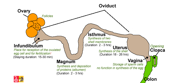
ovulation and are stored in layers in the egg yolk. The mature egg (=
oocyte) is released by the ovary during ovulation and is then incorporated in the funnel-shaped opening (
infundibulum) of the
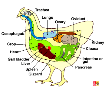
oviduct (More drawings on the anatomy of the hen at a
larger size or
without labels). Fertilization by the sperm of the cock can occur at that location during a short time period (approximately in the first 15 minutes). Therefore, the sperm cells have to pass the whole way through the oviduct from the
cloaca up to the infundibulum. The sperm cells can be stored temporarily in the vagina where selection is taking place. The egg membrane becomes the fertilization membrane in a very short time after penetration by the sperm cell. This membrane inhibits penetration by other sperm cells. The fertilized oocyte (=
zygote) starts immediately with the cleaving process and a small
germ layer is formed at the surface of the yolk. The egg is loaded with albumen at the
magnum. The two semi permeable shell membranes are formed at the
isthmus while the poriferous calcified shell is set off in the uterus. The egg is layed around 24 to 28 hours after fertilization. A light contraction of the egg mass takes place after cooling down and results in the formation of an air chamber between the two aforementioned shell membranes.
For practical courses on
in ovo (in the egg) observations and about later stages of embryonic development see other
website
|
Reproductive organs of the hen and fertilization path
Redrawn and adapted from images on Wikipedia and other photographs |
Early embryology in birds
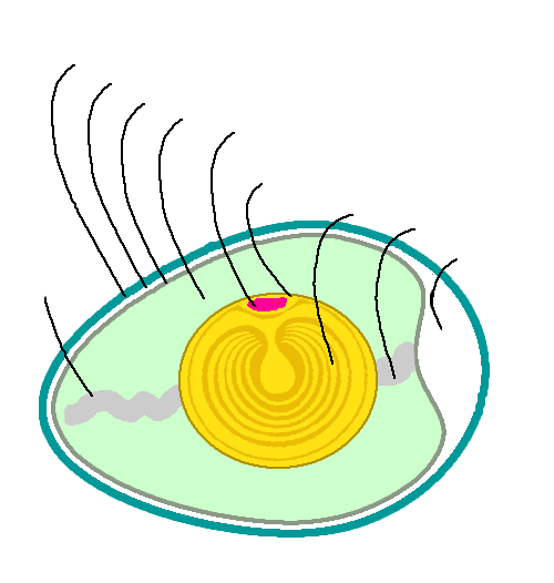
Birds have a
telolecithal type of egg with a large and dense yolk spread throughout most of the egg, but separate from the pole of the developing embryo. The pole of the egg with the lowest concentration of yolk is referred to as the
animal pole, while the opposite part is named the
vegetal pole. Telolecithal eggs undergo discoidal
meroblastic (partial) cleavage (result of cell divisions without increase of mass), where no yolk is incorporated in the embryo. This is not only true for birds, but also for fishes. The embryonic development in birds occurs in the
blastodisc (also called germinal disc or germinal layer).
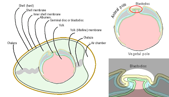 |
Anatomy of a fertilized egg of a chicken; adapted from a Wikipedia image
English name: chicken, Scientific name: Gallus domesticus, Familia: Phasianidae/Meleagridae, Classis: Aves, Phylum: Chordata (subphylum vertebrata), Regnum: Animalia
More drawings on the anatomy of the egg:
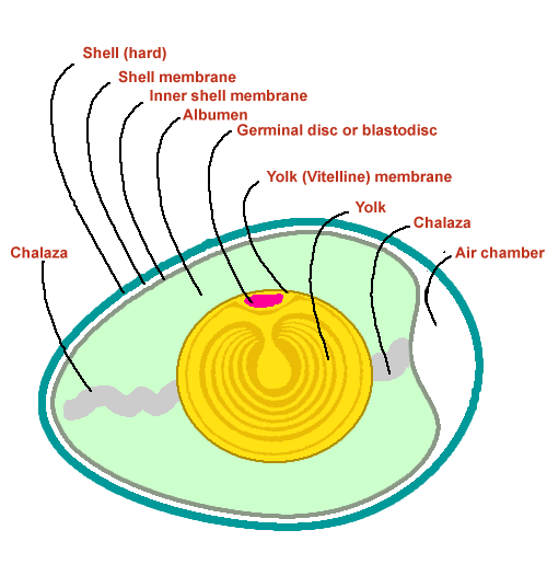 larger size and with more vivid colors (real size 500 x 526 pixels) larger size and with more vivid colors (real size 500 x 526 pixels)
 without labels (real size 500 x 526 pixels)
|
References
Sublime site of the University of New South Wales on embryologie
Figures of Bradley Merrill Patten uit 1920 (The Early Embryology of the Chick. Philadelphia: P. Blakiston's Son and Co.)[/eng 

