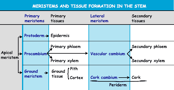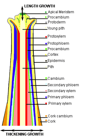
 The apical meristems remain active during the entire development of the plant so that longitudinal extension through cell division as well as cell expansion can occur (see examples here below). In nearly all monocots cell division in primary growth and the consecutive expansion of cells form the only means for the plant to increase in size. During the life of most dicotylous plants, besides primary growth also secondary growth, takes place. This type of growth, called also secondary thickening or lateral growth (lateral = to the side), arises from secondary (newly formed) meristems. 1. From the procambium in the vascular bundles secondary cambium is formed which produces secondary phloem and xylem. 2. In some species cork cambium that makes cork tissue develops from parenchymatic cells in the cortex. Variations in modes of secondary growth are illustrated in the following web pages with two model species: castor bean (POISONOUS PLANT, I.E. THE BEANS ARE EXTREMELY TOXIC!) and Dutchman's pipe.
The cambium (from the Latin word cambiare = to change) is a layer of generative tissue that consists of small thin-walled cells with the capacity to divide. In dicots three types of cambia involved in (secondary) thickening growth can be discerned:
- The fascicular cambium that is present in vessels. In the stem the fascicular tissue forms phloem tissue towards the periphery and xylem tissue to the inside. How fascicular and interfascicular cambium look like and which tissue they generate can be seen from the images in the next web page.
- The interfascicular cambium that has developed from parenchyma cells is located between the -separate- groups of vessels. To the examples.
- The cork cambium that builds layers of cork at the periphery of the bark. More about cork
| Thickening growth: cambium ring versus cambium clusters |
| Diagram of cross-sections through the stem of young and older dicots |
| Closed cambium ring: A > B
| Separate clusters of cambium tissue: C > D |
 |
Example: Castor bean
A Young stage - B Old stage
(A)In the stem of the castor bean a ring of cambium (indicated in green) is present from the beginning of thickening growth. (B) During maturation this cambium layer gives rise to a ring of vascular tissue consisting of xylem (red) to the inside and phloem (blue) to the outside.
| Example: Aristolochia sp.
C Young stage - D Old stage
In young stems of Aristolochia (C) cambium is present in separate clusters in the vessels (fascicular cambium, indicated in green). This fascicular cambium proceeds to the formation of a large amount of secondary tissue (phloem to the outside, indicated in blue and xylem to the inside, indicated in red). Also between the vascular bundles parenchyma cells develop to interfascicular cambium. In older stages (D) arrays of differentiated parenchyma (orange) can be found between the vascular bundles: periclinal divisions occur which give rise to dilatation tissue that compensates for the increase in circumference of the growing stem. |
| Fascicular cambium versus Cork cambium
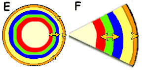
|  | E Fascicular cambium is characterized by bilateral (on two sides) deposition of vascular tissue, namely also here phloem toward the periphery and xylem to the inside.
F Cork cambium, located beneath the epidermis, is involved in cork formation and moreover is mostly active toward the outside.
|


