 |
Chicken: 33-36 h |
At about 33 hours after fertilization, the embryo is about 4 mm long and the first flexion of the originally straight embryo starts in the head region. The cranial flexure will be visible a few hours later. At this stage 12 to 13 somites are formed. The eye vesicles are rather large. The forebrain vesicle or prosencephalon will divide, the midbrain vesicle or mesencephalon remains undivided while the hindbrain vesicle or rhombencephalon will form a series of smaller neuromeres. The sinus rhomboidalis (diamond-shaped) is still present as the only opening of the neural tube and the primitive streak is only rudimentary. The infundibulum (= derived from the diencephalon) appears as a half circular structure at the ventral side of the caudal part of the forebrain. The notochord or chorda dorsalis ends just behind this ventral vesicle. Later on,
36 hours after fertilization, the heart, which has a bilateral origin in the mesodermal layer, is a S-shaped tube which protrudes to the right of the embryo (in upper view). Outside, in the area vasculosa (= forseen of blood vessels) the formation of blood islands continues. The primitive streak can only still be discerned below the sinus rhomboidalis.
Early embryonic developmental features that are shown on this page:
Whole mount preparation 33 hours (More...)
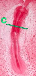
Whole mount preparation 36 hours (More...)
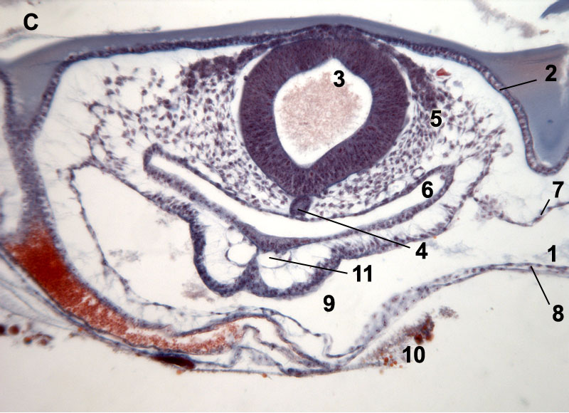
Cross sections 36 hrs; formation of eye, heart and intestines (
More...)
Stage 33 hours
Information:
At about 33 hours after fertilization, the embryo is about 4 mm long and the first flexion of the originally straight embryo starts in the head region and the cranial flexure will be visible a few hours later. At this stage 12 to 13 somites are formed. The eye vesicles are rather large. The forebrain vesicle or prosencephalon will divide, the midbrain vesicle or mesencephalon remains undivided while the hindbrain vesicle or rhombencephalon will form a series of smaller neuromeres. The sinus rhomboidalis (diamond-shaped???) is still present as the only opening of the neural tube and the primitive streak is only rudimentary. The infundibulum (= derived from the diencephalon) appears as a half circular structure at the ventral side of caudal part of the forebrain. The notochord or chorda dorsalis ends just behind this venral vesicle.
| 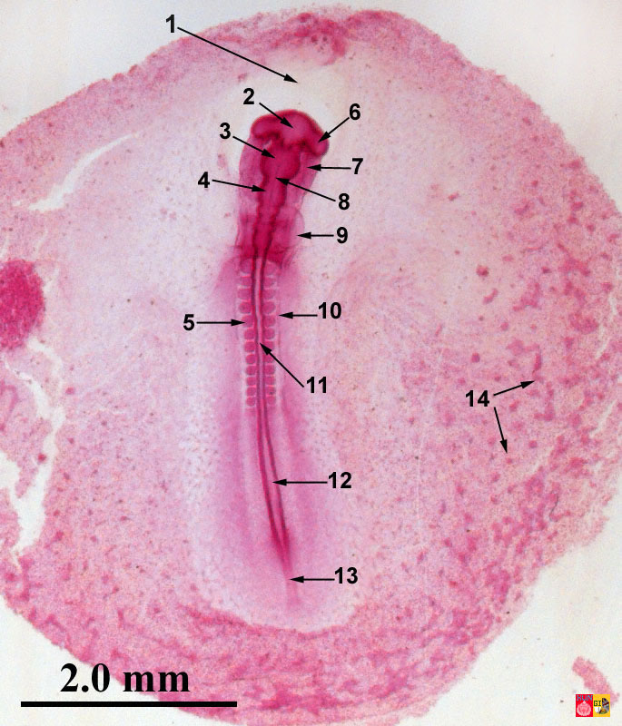 |
Embryology of chicken 33 hours after fertilization: stained whole mout preparation
1 = Proamnion, 2 = Prosencephalon, 3 = Mesencephalon, 4 = Rhombencephalon, 5 = Somite, 6 = Eye vesicle, 7 = Foregut, 8 = Chorda (translucent), 9 = Heart, 10 = Lateral mesoderm, 11 = Spine, 12 = Sinus rhomboidalis, 13 = Primitive streakp, 14 = Blood islands
|
| Dorsalview and longitudinalsection at 33 hrs according to Patten |
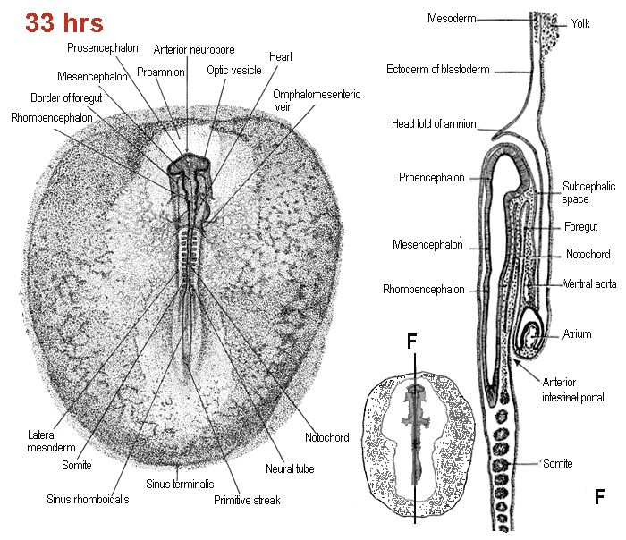 |
| Patten, B.M. (1920). The Early Embryology of the Chick. Philadelphia: P. Blakiston's Son and Co. |
Stage 36 hours
Information:
36-hours after fertilization, the heart is a S-shaped tube which protude to the right of the embryo (in upper view). The further development of the heart is now mesodermal. Outside, in the area vasculosa (= forseen of blood vessels) the formation of blood islands continues. The primitive streak can only still be discerned below the sinus rhomboidalis.
| 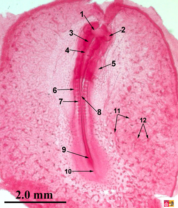 |
Embryology of the chicken 36 hours after fertilization: stained in toto preparation
1 = Prosencephalon, 2 = Eye vesicle, 3 = Mesencephalon, 4 = Rhombencephalon, 5 = Heart, 6 = Lateral mesoderm, 7 = Somite, 8 = Spine, 9 = Sinus rhomboidalis, 10 = Primitive streak, 11 = Small blood vessel, 12 = Blood islands
|
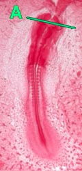
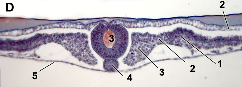
| Cross sections - 36 hours: Formation of eye, heart and intestines |
|
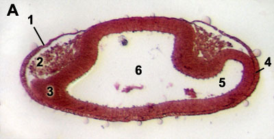
Formation of the eye |
Formation of the eye
1 = (Epidermal) Ectoderm, 2 = Neural crest cells, 3 = Neuroectoderm, 4 = Lens placode, 5 = Cavity of the eye vesicle, 6 = Cavity prosencephalon
|
| 
|
|
Formation of the heart between 25 and 29 hrs (according to Patten but modified) |
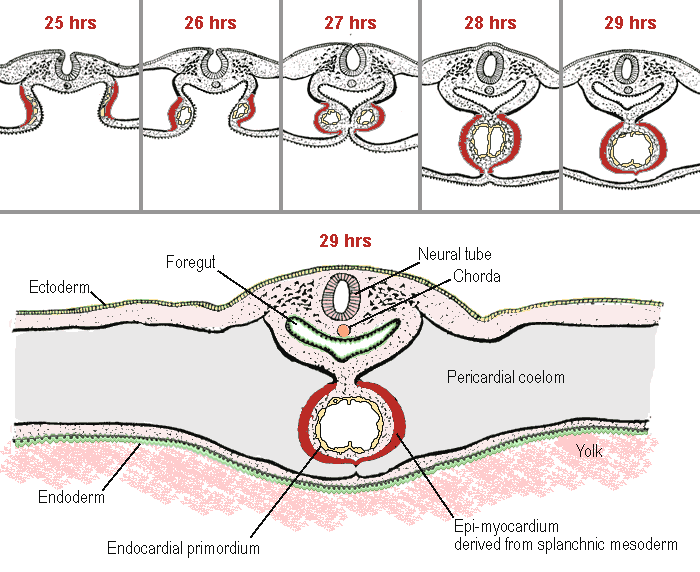 |
| Patten, B.M. (1920). The Early Embryology of the Chick. Philadelphia: P. Blakiston's Son and Co. |
 | 
|  |
|
Heart formation:
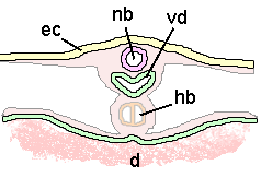
The formation of the heart is mesodermal and arises as two lateral thickenings of the splanchnic mesoderm (primordia) at the level of the head region. These two primordia grow later together in the median plane. Ectoderm (2) and somati mesoderm (4) form the somatopleura; splanchnic mesoderm (5) and endoderm (6) form the splanchnopleura.
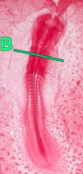
1 = Coelome, 2 = Ectoderm, 3 = Neural tube, 4 = Somatic mesoderm, 5 = Splanchnic mesoderm, 6 = Endoderm, 7 = Foregut, 8 = Heart formation, 9 = (Dorsal) mesocardium, 10 = Head mesenchym, 11 = Chorda, 12 = Endocardium, 13 = Epimyocardium, ec = Ectoderm, nb = Neural tube, vd = Foregut, hb =Heart tube, d = Yolk zoom
|
Intestine port / heart region
1 = Coelome, 2 = (Epidermal) Ectoderm, 3 = Neural tube, 4 = Chorda, 5 = Head mesoderm (Mesenchym), 6 = Foregut, 7 = Somatic mesoderm, 8 = Splanchnic mesoderm, 9 = Pericardial cavity, 10 = Yolk, 11 = Ventral aorta arches zoom |
 |
 |
Neural tube and chorda formation
1 = Lateral plate mesoderm, 2 = Intermediate mesoderm, 3 = Somite mesoderm, 4 = Chorda, 5 = Endoderm zoom |
 |
 |
 last modified: 1 Dec 2011 |
  |
|

|


